MRI (Magnetic Resonance Imaging)
Table of Contents
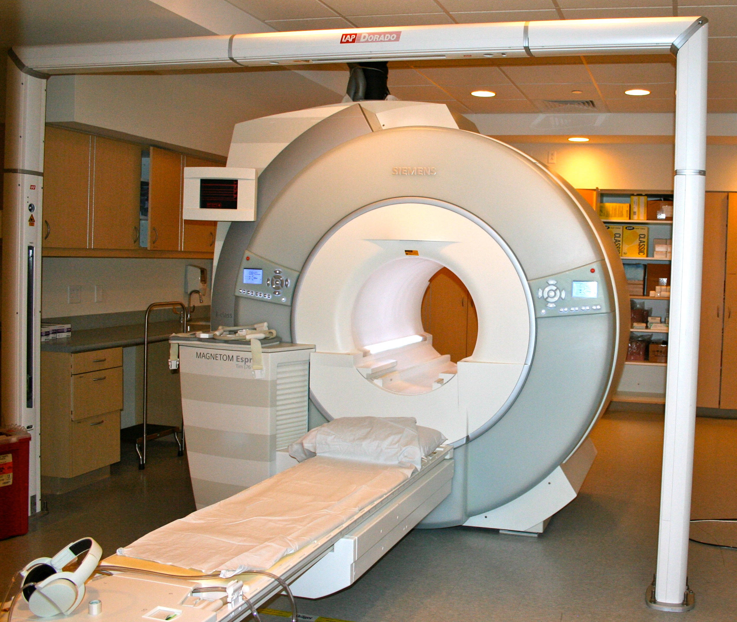
MRI stands for “magnetic resonance imaging” scan. It is a medical test that lets your healthcare provider see detailed images of your internal organs using a large magnet, radio waves, and advanced computer software.
Why would I need an MRI?
MRI scans are used because they give details that cannot be seen in other radiology tests, such as X-rays. MRI scans can help diagnose or look at the size of tumors, see if the tumor can be removed with surgery, and are used to help create the best plan for radiation treatments.
MRI scans used for cancer are most often for:
- Brain and spinal cord tumors.
- Head and neck tumors near the base of the skull.
- Tumors in the chest, near the chest wall or heart.
- Breast cancer patients to check for breast cancer in the other breast.
- Women who have dense breast tissue or are at high risk for cancer.
- Liver cancer.
- Prostate cancer.
MRI scans are most often used for non-emergent imaging tests. This is because MRI scans take longer to be done and read.
How do I prepare for an MRI scan?
Eat normally and take your medication unless your provider tells you differently. Metal can affect your test so you will be asked questions about any metal that could be inside you, like a port, joint replacement parts, internal implants, and so on. Before your MRI, you will be asked to take off any items that may affect the scan, like jewelry, glasses, wigs, dentures, hearing aids, and bras with underwire.
How is an MRI performed?
You will lie flat on a table that moves through a large tube (this is the magnet). The tube can be loud while it is on, and you may be given earplugs or headphones to protect your ears. The test can take anywhere from 30 minutes to 2 hours, based on what areas are being scanned. A radiology technologist will be in a nearby room with a window and microphone to hear and talk to you during the test. You should try to hold still during the test because movement can cause blurry images.
IV contrast may be used during the MRI to see the blood vessels better. This contrast is known as “gadolinium” and is different from the contrast used during a CT scan. If you have an allergy to CT contrast, which is iodine based, there is a good chance you will still be able to have the MRI contrast. Gadolinium cannot be given to anyone with kidney disease because it can harm the kidneys.
What if I am anxious or claustrophobic?
Many people worry about being in a tube during the exam. If you are claustrophobic (afraid of small or confined spaces), your provider may be able to give you medicine to help you relax. You will need to have a ride home after the exam if you have taken medication to relax since you will not be able to drive.
If you are not able to tolerate the MRI after taking medication, an open MRI scan may be an option for you. Open MRI scanners are not as common as standard MRI scanners. The pictures from an open MRI may not be as good, so talk with your provider to see if this is an option for you.
How do I get the results of my MRI?
A radiologist, a doctor that looks at different types of images, looks at the scan and creates a report. The report gives information about normal and abnormal findings. Your provider will be able to talk about your results with you.
If your MRI scan is for radiation planning only, then your radiation oncologist will use the images to build your radiation treatment plan.
