Proton Therapy Delivery: The Equipment
Table of Contents
The equipment required to deliver proton therapy is complex and expensive. The major components of the system are an accelerator, an energy selection system, a beam transport system, a treatment nozzle, and patient positioning and imaging equipment. We will discuss each of these sub-systems:
The Accelerator
Proton therapy requires a proton beam that goes 30-35 cm into the patient’s body. A proton beam capable of this has an energy of about 250 million electron volts (MeV). This means that the proton must pass between two plates that have a voltage of 250 million volts (MV) between the two of them. This is a problem since the highest voltages found on earth are in thunder clouds.
Although these voltages may exceed 250 MV, such voltages cannot be sustained for long. Pretty soon there is a bolt of lightning, and the voltage is discharged (Figure 1a). The maximum manmade voltages of around 30 MV have been attained using Van der Graaf machines. You may remember these from your high school physics class (Figure 1b).
So how do we attain a proton energy of 250 MeV? We accelerate the protons through a much smaller voltage but do it multiple times. The technology for doing this was developed in the early 1930s by Ernest O. Lawrence, a professor at the University of California.
Lawrence wrote down the equation describing the force on a charged particle in a magnetic field that causes it to move in a circle. He wrote down another equation for the mechanical force on a particle moving in a circle. He combined these two equations and realized that there was a resonance condition. If you increase the energy of the particle, it moves to a large radius because it is harder for the magnet to bend it in the circular path. However, the energy increase also means the particle is moving faster. The larger distance it must travel at the new radius is balanced by the increase in speed so that it takes the same time to complete one turn in the magnetic field at the higher energy as it did at the lower energy.
For a particle of given mass and charge and for a known magnetic field, Lawrence could calculate the period of rotation. If he placed a source of protons at the center of the magnet and applied a voltage varying with the appropriate frequency, he could get the proton to gain energy on every rotation moving to a larger radius with each turn. The voltage could be supplied by a radio wave transmitter. Lawrence had invented the cyclotron: he had found a way to accelerate the protons through a small voltage, multiple times.
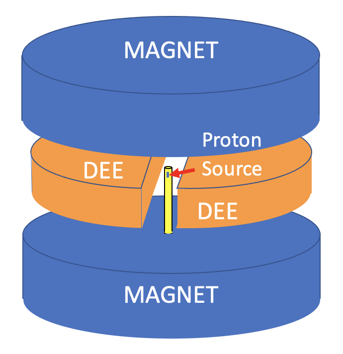
The main components of the cyclotron are a large electromagnet, a source of protons at the center of the magnet, and two semicircular, hollow copper electrodes (known as “Dees”) between the magnet pole pieces. These electrodes are contained in a vacuum enclosure. A simplified diagram of the arrangement is shown below (Figure 2). The vacuum chamber is omitted for clarity.
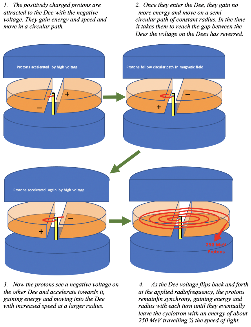
The radiofrequency high voltage is applied between the two Dees. When one Dee has a positive voltage, the other has a negative voltage. These voltages change polarity with the resonance frequency determined by Lawrence’s equations. In a typical cyclotron, this radio frequency is close to FM radio frequencies you may be familiar with, that is around 100 megahertz. This means the voltage on the Dees is changing about every 10 billionth of a second.
Simpler pictures demonstrating the operation of the cyclotron are shown in the cutaway diagrams below (Figure 3):
These diagrams above are an oversimplification. In practice, the voltage on the Dees is typically 50kV and multiple Dees may be used so the protons gain about 200 KeV of energy each turn. Therefore, an energy of 250 MeV takes 1250 turns or 12.5 millionth of a second at a radiofrequency of 100 megahertz. The pole pieces of the magnet are typically 1500 mm in radius so the radius gain each turn is about 1.2 mm.
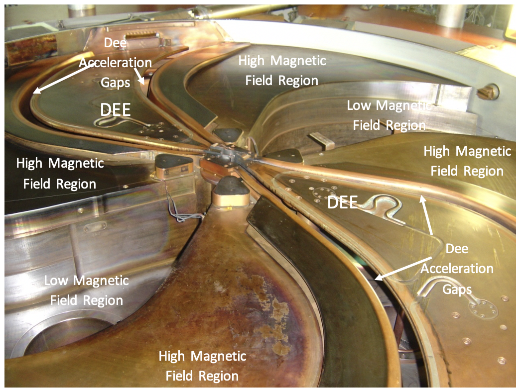
As seen above (Figure 4), the interior of a modern proton therapy cyclotron is complex. There are high and low magnetic field regions where the pole pieces are higher or lower, respectively. In the cyclotron shown here, there are two Dees located in two of the low field areas. In this configuration, there a four Dee gaps where the protons experience acceleration from the radiofrequency voltage.
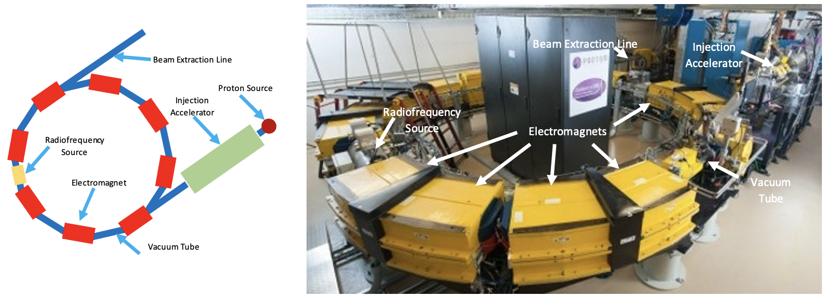
Another type of cyclic accelerator that uses a magnetic field and a radiofrequency to accelerate protons for therapy is the synchrotron (Figure 5). In the synchrotron, the large single magnet of the cyclotron is replaced by a ring of smaller magnets surrounding the vacuum tube, in which the protons travel. The ring has a fixed radius. The magnets are set to give a low magnetic field and low energy protons are injected into the ring from a small accelerator. A radiofrequency source is included in the ring. As the protons circulate, they receive energy from the radio frequency on each turn. To keep them from colliding with the vacuum tube, the magnetic field must be increased on each turn. As the protons are now traveling faster, they arrive at the radio frequency source sooner. Therefore, to maintain the acceleration resonance condition, both the magnetic field and the radiofrequency must be increased in synchrony. The process continues until the desired energy is reached. The energy is determined by the maximum magnetic field attainable and the radius of the vacuum ring, thus, it is possible to control the output energy by selecting the maximum values to which the magnetic field and radio frequency are ramped. This is an advantage over the cyclotron which produces a single output energy.
To achieve an energy of 250 MeV, a ring 6 or 7 meters (24 feet) in diameter is required. The ramping up and down of the magnetic field and radiofrequency takes 1 to 2 seconds and, therefore, the synchrotron produces a pulse of protons every 1 or 2 seconds, unlike a cyclotron that produces a continuous stream of protons.
The exact type of accelerator used to produce the proton beam is not important, the efficacy of the proton beam is not affected. Once the beam is extracted from the accelerator, it passes into the energy selector and/or the beam transport system.
The Energy Selector & Beam Transport System
The Energy Selector
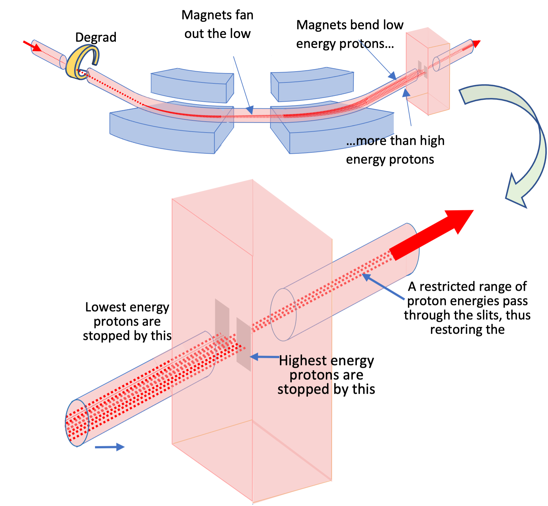
A simplified drawing of the energy selection system used with a cyclotron is shown in Figure 6 below.
A cyclotron produces a single maximum energy. However, since the beam energy determines the depth of penetration of the protons into the body, we need to be able to adjust the beam energy to tailor the beam to the depth of the tumor. To do this, the proton beam from the cyclotron must pass through a degrader and another system of magnets, the “energy selector,” to reduce the energy to that needed for the individual patient’s treatment.
A wedge-shaped piece of graphite is used to reduce the beam energy. The more graphite the beam passes through, the more energy it loses. Unfortunately, in passing through the graphite, the energy spread of the beam is increased and this has an undesirable effect, as it spoils the sharpness of the Bragg peak. To restore the sharpness, the beam is passed through dipole magnets. The low-energy protons in the beam are bent more by the magnetic field than the high-energy protons, so the beam fans out. By placing a slit in the beam path after the dipole magnets, the lowest and highest energy protons can be stopped so that only a limited range of proton energies arrives at the treatment room and the sharpness of the Bragg peak is restored.
As already noted, a synchrotron does not require a degrader/energy selector as the beam energy can be varied during the acceleration process.
Beam Transport System
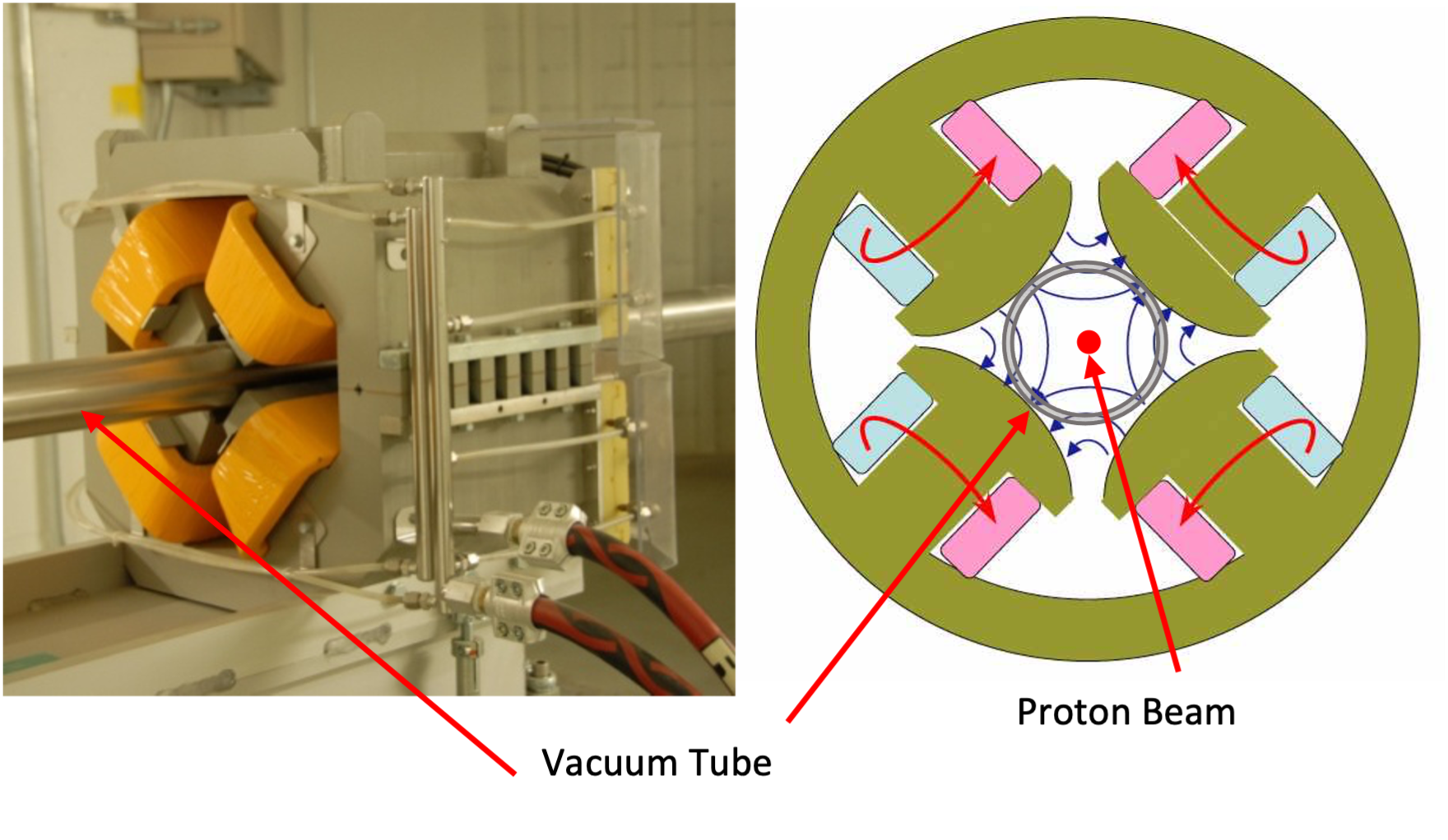
Once the beam leaves the synchrotron or the energy selection system, it must be transported to the treatment room. In a multiroom system, this may involve transporting the room through a vacuum tube up to 160 feet in length. As the proton beam travels through the tube, it diverges and must be focused. Special electromagnets with two pairs of diametrically opposed pole pieces, known as quadrupole magnets (Figure 7), keep the beam focused in a manner similar to the way a lens is used to focus a light beam.
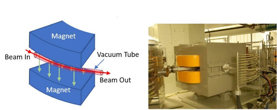
More electromagnets are used to steer the beam into the treatment room. These are dipole magnets that bend the beam (Figure 8).
The Gantry
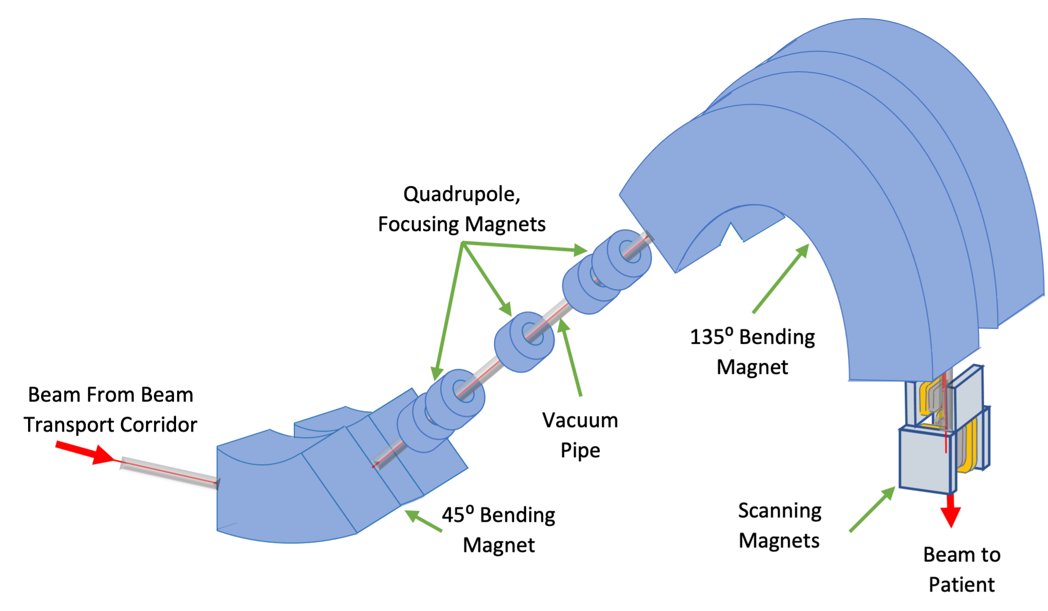
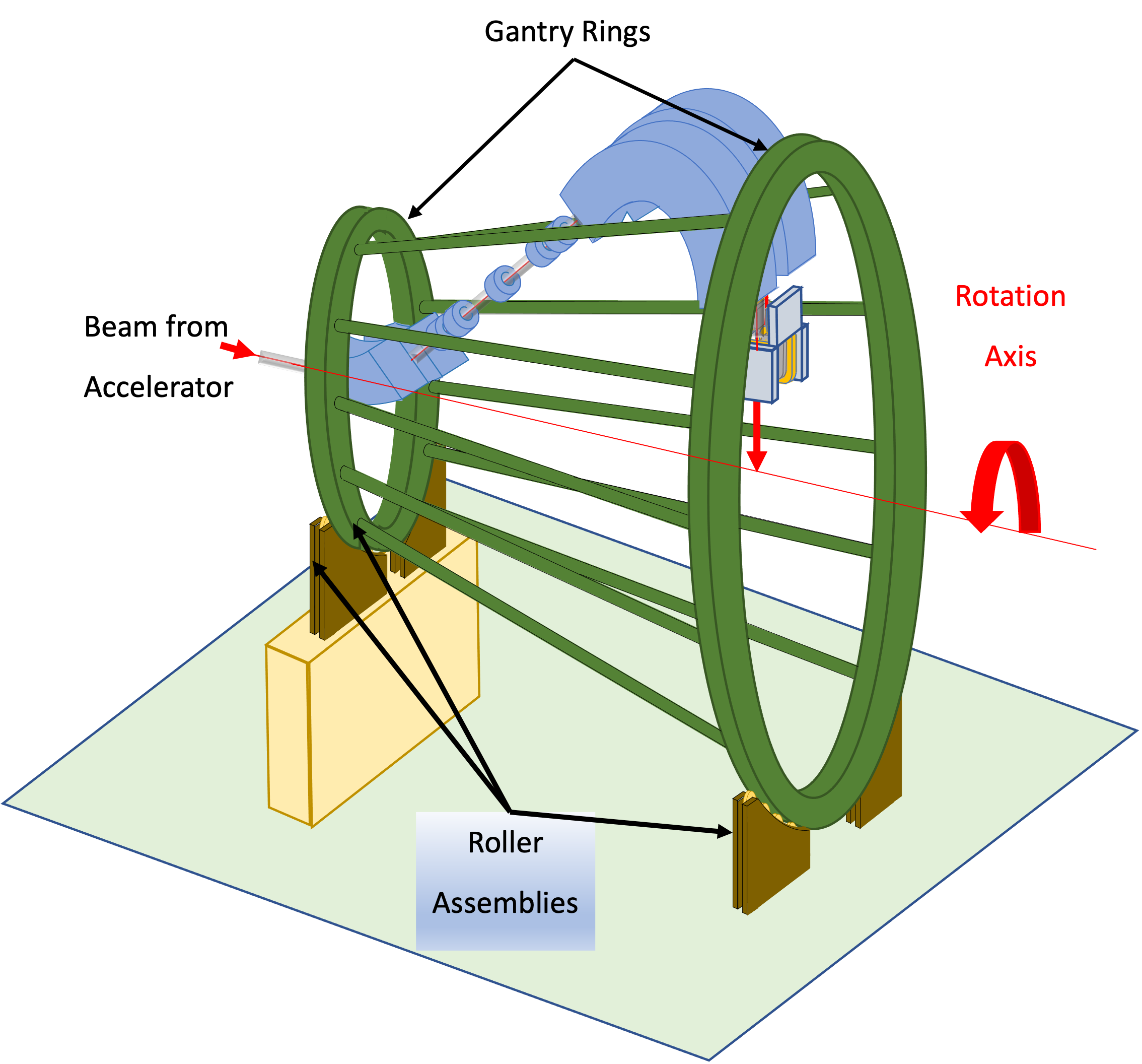
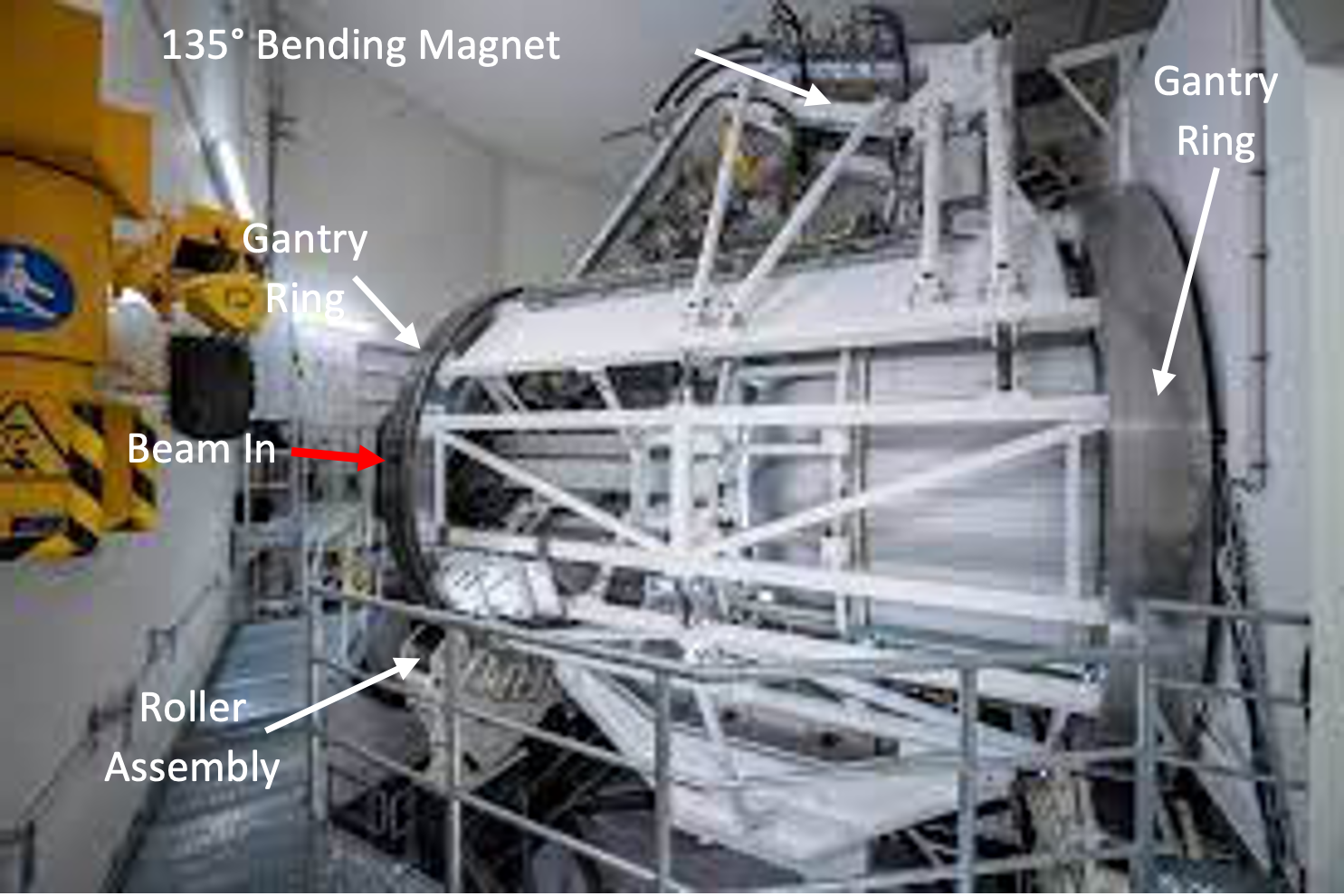
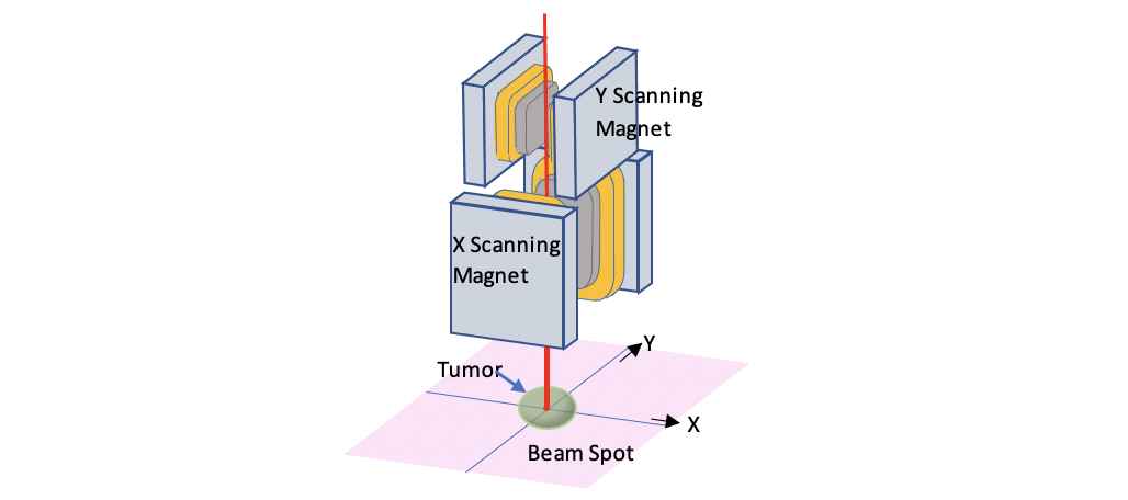
Once the beam leaves the beam transport corridor and enters the treatment room, it needs to be precisely delivered to the patient’s tumor. The ideal way is to rotate the beam 360⁰ around the tumor. To do this, more bending magnets and focusing magnets (Figure 9) are mounted on a gantry (Figures 10 and 11) that can rotate around the patient. A pair of dipole magnets, the scanning magnets, are used to steer the tumor.
In the Treatment Room
The Treatment Nozzle
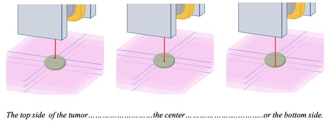
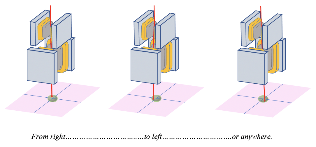
Once the beam is directed towards the patient’s tumor by the 135⁰ bending magnet, it passes into the treatment “nozzle.” This contains the scanning magnets and instrumentation for monitoring the dose and the beam position. The proton beam’s Bragg peak is about 1 cm in width and the proton beam itself is about 1 cm in diameter, so the beam is delivered as a series of 1 cm diameter spheres or beam “spots.” Many of these small spots must be delivered to the tumor to provide uniform dose coverage (Figures 12, 13, and 14). The scanning magnets are used to steer the beam spot across the tumor volume.
An average-sized tumor requires as many as 400 spots in each layer and 20 layers to give excellent dose coverage. These 8000 spots are typically delivered in less than one minute.
Patient Positioning and Imaging
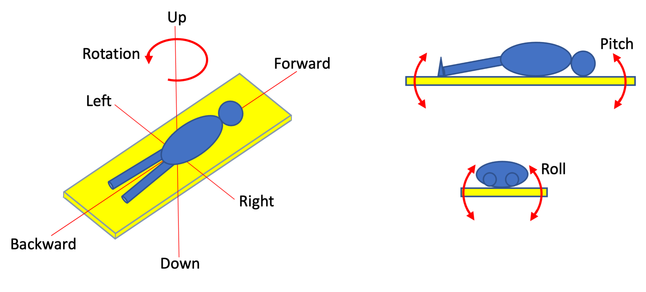
The final step in preparing for a patient’s treatment is to align the patient accurately in front of the proton beam. Two pieces of equipment are critical in achieving this: The patient treatment couch and the x-ray imaging equipment in the treatment room. Prior to treatment the patient undergoes a simulation procedure, during which multiple CT x-ray images are taken of the patient in the treatment position and marks are made on the patient’s skin so they may be repositioned in front of the proton beam. Later, after all the treatment planning has been completed, the patient lies on the computer-controlled couch and its position is adjusted to align the marks made on the patient’s skin with lasers that define the beam position. To do this, the couch can be moved in many directions (Figure 15); up and down, backwards and forwards, left and right, it can be rotated, it can even be tilted from head to toe (pitch) or left to and right (roll), just like the pitch and roll of a boat. Some couches use robots similar to those used in the manufacturing industry (Figure 16).
The last step in preparing the patient for treatment is to confirm the couch position and patient alignment by taking x-ray images of the patient in the treatment position. These images are then compared to the images taken earlier during the simulation procedure. If the images agree, the treatment proceeds, otherwise there are more couch position adjustments followed by x-ray confirmation. The imaging systems vary but typically they are attached to the gantry (Figure 17).
All this proton therapy equipment is linked together and controlled by computers to ensure that the treatment can be delivered safely and with millimeter precision.

